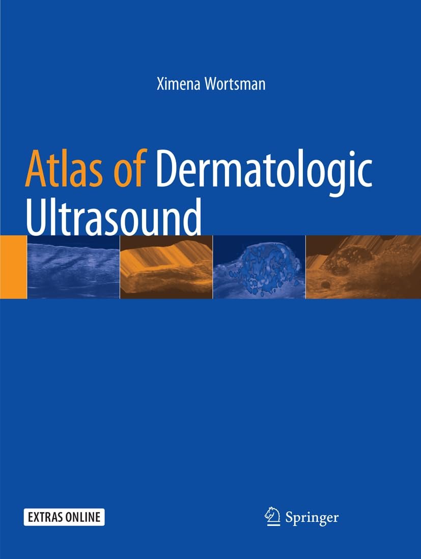
The “Atlas of Dermatologic Ultrasound” is a comprehensive visual reference that showcases the utility of ultrasound in dermatology. This atlas provides detailed insights into the use of ultrasound imaging for diagnosing and managing various dermatological conditions.
Authored by experts in the field, the atlas features high-quality ultrasound images accompanied by explanatory text, offering readers a thorough understanding of the diagnostic capabilities of dermatologic ultrasound. From common skin disorders to more rare and complex conditions, the atlas covers a wide spectrum of dermatological pathologies.
Key features of the “Atlas of Dermatologic Ultrasound” may include:
- Comprehensive Coverage: The atlas covers a wide range of dermatological conditions, illustrating how ultrasound can be used for diagnosis, assessment, and treatment monitoring.
- High-Quality Images: The atlas includes high-resolution ultrasound images that provide detailed visualization of skin structures, lesions, and underlying tissues.
- Clinical Correlation: Each image is accompanied by explanatory text that correlates ultrasound findings with clinical features, aiding in accurate diagnosis and management.
- Diagnostic Techniques: The atlas may also include descriptions of ultrasound techniques used in dermatology, such as high-frequency ultrasound, Doppler imaging, and elastography.
- Educational Resource: Whether for dermatologists, radiologists, or other healthcare professionals, the atlas serves as an educational resource for understanding the role of ultrasound in dermatology.
Overall, the “Atlas of Dermatologic Ultrasound” is a valuable tool for healthcare professionals seeking to enhance their diagnostic skills and improve patient care in dermatology.

 Anaesthesia books
 Behavioral Science Books
 Cardiology Books
 Obstetric and Gynecology
 AMC Books
 Prepladder Notes
 Stethoscope
 Dermatology Books
 Neurosurgery Books
 Dentistry Books
 ENT Books
 Anatomy Books
 Biochemistry Books
 Biostatistics Books
 Plab Books
 Radiology Books
 Surgery Books


Reviews
There are no reviews yet.